Home
/ Graafian Follicle Histology, Ovarian Follicle Wikipedia, Should be differentiated from cystic granulosa cell tumor.
Graafian Follicle Histology, Ovarian Follicle Wikipedia, Should be differentiated from cystic granulosa cell tumor.
Graafian Follicle Histology, Ovarian Follicle Wikipedia, Should be differentiated from cystic granulosa cell tumor.. Graafian follicles can be defined structurally as a heterogeneous family of relatively large follicles (400 µm to >2 cm at ovulation) that display an antrum containing follicular fluid, or liquor folliculi. (redirected from graafian follicle) an ovarian follicle is a roughly spheroid cellular aggregation set found in the ovaries. The graafian follicle is the stage after the first meiotic division has completed but before ovulation. Follicles, classified as primordial, primary, secondary and graafian (mature), house developing germ cells (oocytes) and secrete estrogen. It is as if the follicle was a water filled basketball, and the ovum was a ping pong ball, wrapped in paper and then glued to the
The oocyte is surrounded by a layer of follicular cells, which remain. Start studying histology of the reproductive system. What are the components of a mature/preovulatory/graafian follicle? The antrum is a characteristic structural feature of all graafian follicles. (redirected from graafian follicle) an ovarian follicle is a roughly spheroid cellular aggregation set found in the ovaries.
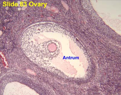
Histology Laboratory Manual from www.columbia.edu The secondary oocyte, having undergone the first meiotic division, is located eccentrically. The graafian follicle is the stage after the first meiotic division has completed but before ovulation. Follicles, classified as primordial, primary, secondary and graafian (mature), house developing germ cells (oocytes) and secrete estrogen. The graafian, or mature, follicle may be up to 2.5 cm in diameter at the time of ovulation, and it protrudes from the surface of the ovary. The oocyte is now a 2n haploid. The follicle is characterized by a large follicular antrum that makes up most of the follicle. The follicle is characterized by a large follicular antrum that makes up most of the follicle. Fluorescence microscopy, colored with dapi.
When this happens, the primary follicle has matured into a secondary follicle.
May occur at any age, with a variable clinical presentation depending on age and etiology. It therefore contains a 2n haploid oocyte. An outer layer of theca cells and granulosa cells surround a vesicle containing fluid and the oocyte. Once the structure is selected you can go to the related images to see if there are more views of the selected structure. The follicular cyst of ovary (or ovarian functional cyst) is a type of functional simple cyst, and is the most common type of ovarian cyst. The graafian follicle is the stage after the first meiotic division has completed but before ovulation. The first meiotic division is now completed, and the oocyte is now a secondary oocyte, and starts its second meiotic division. Graafian follicle, 25x the final stage of development is the graafian or mature follicle. Benign cyst lined by an inner layer of granulosa cells with an outer layer of theca cells. This oocyte release is called ovulation, and it happens around the midpoint of the cycle (about day 14). This image is a histological section of a mature follicle within the ovary. Graafian follicles can be defined structurally as a heterogeneous family of relatively large follicles (400 µm to >2 cm at ovulation) that display an antrum containing follicular fluid, or liquor folliculi. The secondary oocyte, having undergone the first meiotic division, is located eccentrically.
The secondary oocyte, having undergone the first meiotic division, is located eccentrically. With continued development, the follicle becomes a graafian or ovulatory follicle view image (this follicle is actually rather small to be a real graafian follicle). The diminished cumulus oophoros surrounding the oocyte is called the corona radiata. May occur at any age, with a variable clinical presentation depending on age and etiology. One type of simple cyst, which is the most common type of ovarian cyst, is the follicular cyst of ovary, or graafian follicle cyst, or follicular cyst.
Female Reproductive System Quiz Slide 1 Answer from education.med.nyu.edu The graafian follicle is the stage after the first meiotic division has completed but before ovulation. Fine, now learn the details histology of mature or graafian follicle from animal ovary. The diminished cumulus oophoros surrounding the oocyte is called the corona radiata. It secretes hormones that influence stages of the menstrual cycle. The oocyte is now a 2n haploid. Tertiary follicle or graafian follicle. Zona pellucida is very conspicuous. The graafian, or mature, follicle may be up to 2.5 cm in diameter at the time of ovulation, and it protrudes from the surface of the ovary.
The arrow bar shows the diameter of one mature or graafian follicle.
The antrum is a characteristic structural feature of all graafian follicles. An outer layer of theca cells and granulosa cells surround a vesicle containing fluid and the oocyte. After it releases its oocyte, the graafian follicle is called the corpus luteum. The precise steering mechanism that leads to the selection of a follicle and its subsequent maturation to become a graafian follicle is still unknown. An antral follicle (or graafian follicle) is an ovarian follicle during a certain latter stage of folliculogenesis. Measures ≥ 3 cm, as opposed to cystic follicle, which measures < 3 cm. It secretes hormones that influence stages of the menstrual cycle. One type of simple cyst, which is the most common type of ovarian cyst, is the follicular cyst of ovary, or graafian follicle cyst, or follicular cyst. Women begin puberty with about 400,000 follicles, each with the potential to release an egg cell (ovum) at ovulation for fertilization. Graafian follicle, 25x the final stage of development is the graafian or mature follicle. Should be differentiated from cystic granulosa cell tumor. The first meiotic division is now completed, and the oocyte is now a secondary oocyte, and starts its second meiotic division. Mature (graafian) follicle the mature (graafian) follicle is characterized by an enlarged follicular antrum and thinned granulosa layer.
Before ovulation a growth spurt of the tertiary follicles takes place. Fluorescence microscopy, colored with dapi. The first meiotic division is now completed, and the oocyte is now a secondary oocyte, and starts its second meiotic division. 1 o oocyte is surrounded by a layer of granulosa cells (the corona radiata) and rests on a small mound of granulosa cells called cumulus oophorus. The oocyte is surrounded by granulosa cells, which secrete fluid into the cavity of the follicle.
Female Reproductive from www.ouhsc.edu Lh causes the graafian follicle to move to the surface of the ovary and release its little oocyte into the world. Fluorescence microscopy, colored with dapi. The secondary oocyte, having undergone the first meiotic division, is located eccentrically. The antrum is a characteristic structural feature of all graafian follicles. An antral follicle (or graafian follicle) is an ovarian follicle during a certain latter stage of folliculogenesis. (redirected from graafian follicle) an ovarian follicle is a roughly spheroid cellular aggregation set found in the ovaries. After the first meiotic division, most of the cytoplasm goes into one of the two daughter cells. The oocyte is surrounded by granulosa cells, which secrete fluid into the cavity of the follicle.
The graafian follicle is the follicular stage after the first meiotic division but before ovulation.
Characterized by a large fluid filled antral cavity surrounded by compressed granulose cells. Zona pellucida is very conspicuous. In this lecture i discuss the various stages of ovary development. Once the structure is selected you can go to the related images to see if there are more views of the selected structure. The secondary oocyte, having undergone the first meiotic division, is located eccentrically. The secondary oocyte, having undergone the first meiotic division, is located eccentrically. One type of simple cyst, which is the most common type of ovarian cyst, is the follicular cyst of ovary, or graafian follicle cyst, or follicular cyst. The second division then starts, and a graafian follicle is formed. The first meiotic division is now completed, and the oocyte is now a secondary oocyte, and starts its second meiotic division. Fine, now learn the details histology of mature or graafian follicle from animal ovary. With continued development, the follicle becomes a graafian or ovulatory follicle view image (this follicle is actually rather small to be a real graafian follicle). Using your mouse, select a histological structure to learn more about it. (redirected from graafian follicle) an ovarian follicle is a roughly spheroid cellular aggregation set found in the ovaries.